
The three main research/educational areas of the Surgical Neuroanatomy Lab are: endoscopic skull base anatomy, microsurgical neuroanatomy, and white matter anatomy/brain connectivity/surgical planning.
Endoscopic Skull Base Anatomy
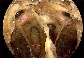
The Endoscopic Endonasal Approach (EEA) has revolutionized skull base neurosurgery. The EEA has anatomical and technical advantages over open skull base approaches for the treatment of selected lesions. EEA is not minimally invasive but maximally effective for the treatment of a wide variety of skull base lesions. The Surgical Neuroanatomy Laboratory at the University of Pittsburgh has pioneered anatomical work on the area of skull base endoscopy, and its goal is to continue providing landmark contributions to the skull base community. Meticulous knowledge of the ventral skull base anatomy as seen from the endoscopic perspective is critical to apply endonasal endoscopic surgery in an effective and safe manner.
Microsurgical Neuroanatomy
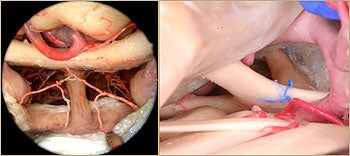
Conventional skull base approaches are being compared with novel endoscopic endonasal approaches to aid in understanding indications and limitations of different but complementary skull base approaches. Contemporary skull base surgeons should combine expertise in open and endoscopic skull base approaches to select the most appropriate approach and technique for each particular case. Emphasis is made on the circumferential conceptualization of the skull base and the selection of “anatomically-favorable” surgical routes.
White Matter Anatomy
Dissection of the white matter fiber tracts provides a unique insight into the complex intrinsic architecture of the brain and builds up an essential knowledge for operating on intra-axial tumors. A unique feature of our white matter studies is the combination with advanced imaging techniques, such as High-Definition Fiber Tractography (HDFT), to facilitate greater understanding of brain connectivity “in-vivo” and in neurosurgery patients.